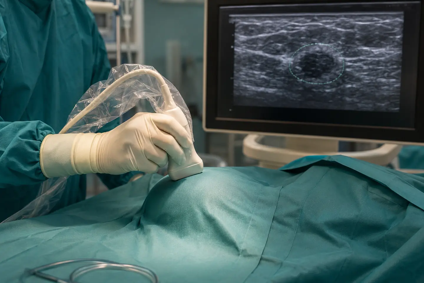
Intraoperative ultrasound means using an ultrasound probe in the operating theatre to see a breast lesion and surrounding tissue in real time. It can connect preoperative imaging with what the surgeon sees and feels during the operation. The technique is particularly useful for breast-conserving surgery when the target is visible on ultrasound.
Localising the lesion accurately
Some breast cancers and benign lesions are easy to feel; others are small, deep or non-palpable. If a lesion is clearly visible on ultrasound, the surgeon can scan before the incision, confirm its position, measure its depth and plan the shortest appropriate route. During removal, repeated views help keep the target centred within the specimen.
This can reduce dependence on touch alone. It may also complement or, in selected ultrasound-visible cases, avoid a separate wire-localisation procedure. Lesions seen only on mammography or MRI still need another localisation method.
Helping achieve clear margins
Breast-conserving surgery aims to remove the cancer with an adequate rim of tissue while preserving as much healthy breast as safely possible. Removing too little can leave an involved margin and lead to another operation; removing too much can affect breast shape.
Real-time ultrasound shows the relationship between the lesion and planned edges. If one margin looks close, the surgeon may remove an additional directed piece during the same operation. In the COBALT randomised trial of palpable, ultrasound-visible breast cancers, ultrasound-guided surgery reduced involved margins and removed smaller volumes than palpation-guided surgery.
Examining the specimen in theatre
After excision, the removed tissue can be scanned to confirm that the imaging target is inside and to estimate its distance from the specimen edges. The specimen is oriented for pathology so each margin can be identified. This immediate feedback can guide further excision while the patient is still anaesthetised.
Ultrasound does not see microscopic cancer cells. The final margin result comes from the pathology laboratory, and a second operation can still be necessary.
Other uses during breast procedures
Intraoperative ultrasound can help guide drainage of a deep collection, identify a clip or marker in selected cases, assess anatomy around a lesion and support planning of an oncoplastic resection. Ultrasound is also used in other perioperative tasks such as regional anaesthetic blocks, but these are different applications with different training requirements.
For aesthetic or implant surgery, ultrasound may help assess a pre-existing implant or fluid collection in selected situations, but it is not a routine substitute for careful surgical technique or formal diagnostic imaging.
Who benefits most?
The main requirement is that the target can be seen reliably on ultrasound and that the operating surgeon or imaging professional is trained to use the technique. It is most valuable when localisation influences incision, specimen volume or margin direction. Very diffuse disease, microcalcifications without an ultrasound correlate or lesions seen only on MRI need other strategies.
The decision is made during preoperative review of images. Marking the patient and scanning before theatre can confirm that the planned method is feasible.
Limits and quality safeguards
Ultrasound is operator dependent. Probe pressure, orientation and image interpretation can affect apparent dimensions. Sterile technique, appropriate equipment, specimen orientation and communication with radiology and pathology are essential. A normal-looking ultrasound edge is not a guarantee of a clear microscopic margin.
The practical value
In the right case, intraoperative ultrasound gives the breast surgeon immediate visual information rather than relying only on preoperative images and touch. It can support accurate localisation, tissue preservation and margin-directed decisions. Its value is greatest as one part of consultant-led, multidisciplinary planning—not as a replacement for pathology or oncological principles.
Sources reviewed
PMC — COBALT randomised controlled trial of ultrasound-guided surgery: https://pmc.ncbi.nlm.nih.gov/articles/PMC4695494/
PMC — Meta-analysis of intraoperative ultrasound-guided breast-conserving surgery: https://pmc.ncbi.nlm.nih.gov/articles/PMC3779206/
PMC — Intraoperative ultrasound and surgical margins in breast cancer: https://pmc.ncbi.nlm.nih.gov/articles/PMC9750786/