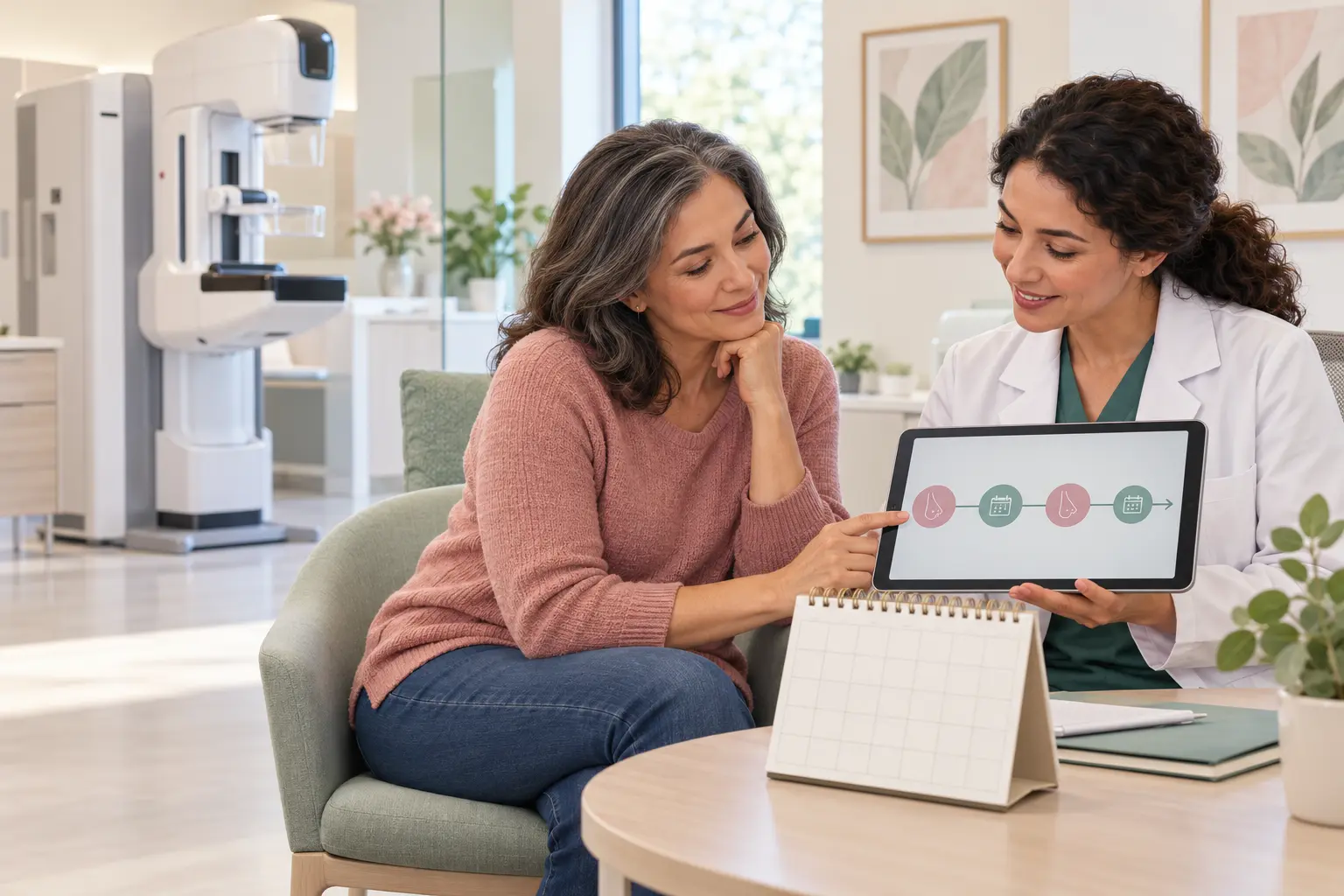
Seeing “calcifications” in a mammogram report can be worrying. The useful next question is not simply whether calcium is present, but what the radiologist recommends after assessing its appearance and comparing earlier images. This guide concerns a newly assessed imaging finding—not the routine surveillance timetable after cancer treatment.
What are breast calcifications?
These are calcium deposits seen on mammography, usually too small to feel. Larger deposits are commonly benign; tiny deposits can also be benign, although some patterns raise concern for ductal carcinoma in situ (DCIS) or invasive cancer. They are not caused by calcium in your diet. The word “microcalcifications” alone does not establish cancer. National Cancer Institute: mammograms
Why are extra views requested?
Magnified mammogram views help the radiologist examine the deposits and their extent. Some calcifications have clearly benign features; others need tissue sampling. A recall is an assessment step, not a cancer diagnosis. NHS breast-screening assessment guidance, updated January 2026
Bring previous images if the centre does not already have them. Ask for the final assessment category and a written recommendation rather than trying to judge a photograph of the scan yourself.
How does BI-RADS guide the next step?
BI-RADS is an imaging assessment system, not a cancer stage. Category 0 means assessment is incomplete. Category 2 describes a benign finding and usually means routine screening. Category 3 means probably benign, with short-interval imaging—often starting at six months. Categories 4 and 5 generally lead to a biopsy recommendation; tissue analysis establishes the diagnosis. Follow the date and action in your own report. American Cancer Society: understanding your mammogram report
Do not independently substitute a follow-up scan for a recommended biopsy. Ask the breast team to explain any uncertainty and the alternatives before deciding.
Why might a normal ultrasound not settle the question?
Some mammographic calcifications are not visible on ultrasound. In that situation, mammography-guided stereotactic biopsy can target the finding. It commonly uses local anaesthetic and a vacuum-assisted needle to collect tissue; a small marker may be left to identify the sampled location. It is a diagnostic procedure, not automatically a cancer operation. ACR/RSNA: stereotactic breast biopsy
Tell the team about possible pregnancy, allergies, medicines and supplements. Ask for a personalised medication plan; do not stop a prescribed blood thinner on your own.
How does the team know the correct area was sampled?
When suspicious calcifications are biopsied, an x-ray of the samples checks that representative deposits were obtained. The tissue result must then explain the imaging finding. If the target was not adequately sampled, repeat image-guided sampling or another diagnostic approach may be needed. NHS screening guidance: calcification assessment
For the separate issue of mismatched results, read a benign biopsy that does not explain suspicious imaging. Do not infer that every benign result means the same follow-up plan; ask what the precise diagnosis means for you.
Leave with a clear results and follow-up plan
Ask: “Which breast and area are being assessed, and what is the final imaging category?”
Ask: “If a biopsy is recommended, which imaging method will guide it, and how will adequate sampling be checked?”
Ask: “Who will explain the pathology result and confirm whether it matches the scan?”
Ask: “What is my next appointment date, and whom should I contact if the result or appointment does not arrive?”
Keep the report, image-access details and written plan together. For people already treated for cancer, our post-treatment mammography guide answers a different question about ongoing surveillance.
Symptoms that should not wait
Report a new breast or armpit lump, persistent skin change, new nipple inversion or non-milk discharge promptly—even after reassuring imaging. These symptoms need assessment, but do not by themselves prove cancer. NCI: breast symptoms
After biopsy, seek same-day advice for worsening pain, fever, increasing redness or swelling, or bleeding or fluid from the wound. Follow the centre's dressing and activity instructions. Seek emergency care for uncontrolled heavy bleeding, collapse or severe breathing difficulty. NHS: biopsy aftercare and urgent symptoms
Choosing a breast-assessment team in Cairo
When searching for “best breast surgeon in Cairo”, compare verifiable registration, breast-specific experience, access to specialist radiology and pathology, documented sample–image agreement, and reliable results follow-up. The phrase is not an objective ranking. Read our objective surgeon-selection guide.
Educational scope and references
This is general education, not personal medical advice or an interpretation of your mammogram. Your breast team must review the images, symptoms, history and any pathology before recommending follow-up or treatment. Sources checked on 8 November 2026; six direct official or professional references are linked here and listed below. No individual medical-review claim is made for this article.