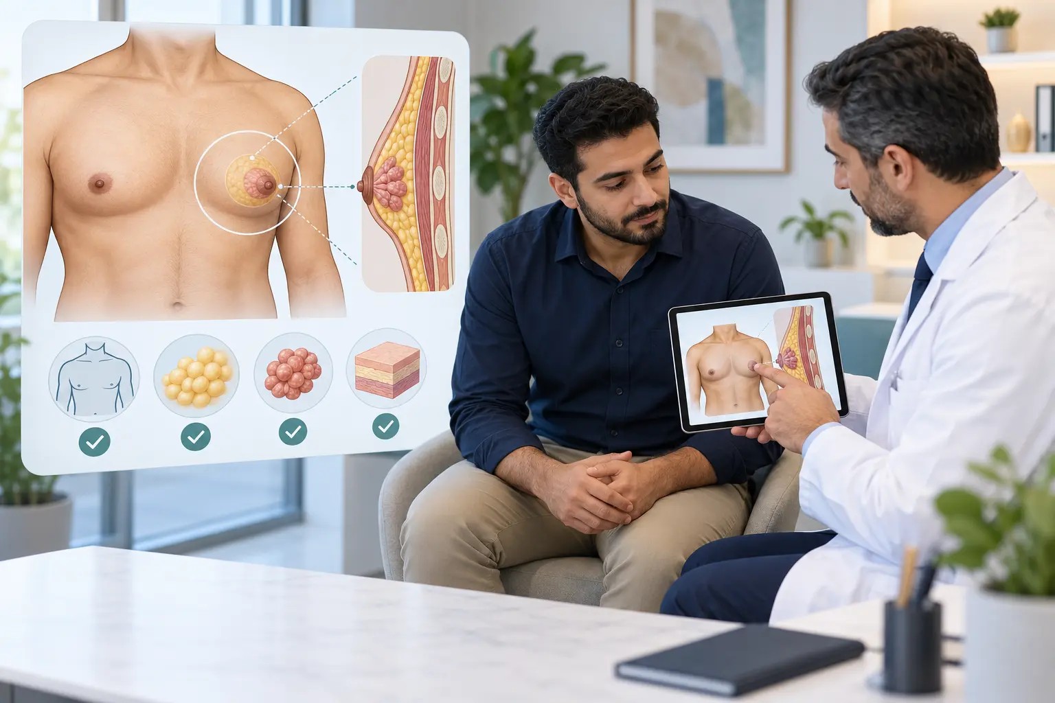
The correct imaging plan after breast-cancer surgery depends on how much breast tissue remains, whether radiotherapy was given, the type of reconstruction, age, inherited risk and the findings from treatment. A routine surveillance mammogram is different from diagnostic imaging for a new symptom. This guide helps you understand the questions to settle with your own breast team.
What is surveillance mammography?
Surveillance mammography is planned imaging after treatment to look for a new cancer or a recurrence in breast tissue that remains. It does not replace clinical review or assessment of a new lump, skin change or other symptom.
The ASCO follow-up guideline published in October 2026 recommends regular history, physical examination and mammography after primary breast-cancer treatment. It also supports a risk-based approach because some people need closer follow-up than others. ASCO breast-cancer follow-up and surveillance guideline, 2026
After breast-conserving surgery
After lumpectomy or another breast-conserving operation, breast tissue remains on the treated side, so mammography usually continues for both breasts. The American Cancer Society states that most experts recommend a mammogram of the treated breast about 6–12 months after treatment ends, followed by mammography at least yearly. Surgery and radiotherapy can change how the breast looks on imaging; the first post-treatment study helps form a new baseline. American Cancer Society: mammograms after breast-cancer surgery
The exact first date should come from the treating team. It can differ when radiotherapy is still in progress, a wound has not healed, extra surgery is planned or an earlier diagnostic study is needed. For the operation itself, see breast-conserving surgery explained and the separate guide to lumpectomy margins.
After a mastectomy
After removal of one breast, routine screening mammography is generally not performed on the mastectomy side, while mammography normally continues for the remaining breast. NICE specifically advises against mammography of the ipsilateral soft tissues after mastectomy. NICE NG101 follow-up imaging recommendations
After bilateral mastectomy, there may be too little breast tissue for routine mammography. This does not mean follow-up has ended: the skin, chest wall, scars and lymph-node areas still need symptom-led clinical assessment. Do not wait for a future scan if you feel a new lump or notice a persistent change.
Do nipple-sparing surgery and reconstruction change the answer?
They can. Nipple-sparing mastectomy may leave a small amount of tissue under the nipple, so the treating team should document whether any imaging is planned. Read the separate guide to nipple-sparing mastectomy.
Screening and diagnostic imaging are also different after reconstruction. The ACR rates screening mammography as usually not appropriate after mastectomy with no reconstruction, while it may be appropriate in selected people with autologous reconstruction. A new lump or other concern in a reconstructed breast can still require targeted ultrasound, diagnostic mammography or MRI. ACR Appropriateness Criteria: imaging after mastectomy and reconstruction
Therefore, do not assume that “reconstruction” automatically means either lifelong mammograms or no imaging. Ask which operation and reconstruction you had, what tissue remains and what the written surveillance plan says. For planning differences, see immediate versus delayed reconstruction.
Are ultrasound, MRI, CT, PET or blood tests routine?
Not for every person without symptoms. NICE does not recommend routine ultrasound or MRI for post-treatment surveillance after early invasive breast cancer or DCIS. MRI can still be appropriate for a separate high-risk indication or a specific clinical or imaging question. CT, PET, bone scans and blood tests are used when the history, examination or individual risk creates a reason—not simply as a substitute for mammography.
In the randomized GIVIO trial, 1,320 women received either intensive scheduled testing with bone scans, liver ultrasound, chest X-rays and laboratory tests or clinically indicated testing; both groups had yearly mammography. The intensive programme did not improve survival or health-related quality of life. The study is old, but it remains foundational evidence behind avoiding indiscriminate whole-body surveillance in people without symptoms. GIVIO randomized trial
How should you prepare for the first post-treatment mammogram?
- Confirm whether the appointment is routine surveillance or investigation of a symptom. - Bring or arrange access to previous mammograms, ultrasound, MRI and operative details when the imaging centre does not already hold them. - Tell the radiographer which breast was treated, the surgery and radiotherapy dates, and whether you have an implant or flap reconstruction. - Mention focal pain, a new lump, skin change or limited shoulder movement before positioning begins. - Ask who will issue the report, when to expect it and who will act on an abnormal or incomplete result.
Postoperative tenderness can make positioning uncomfortable. Tell the radiographer rather than silently enduring severe pain; the team can adapt positioning or decide whether the examination should be rescheduled.
What if you are recalled after the mammogram?
A recall is not the same as a cancer diagnosis. Postoperative scar tissue, fat necrosis, calcifications and an incomplete comparison with old images can all require extra views or ultrasound. If a finding remains uncertain, image-guided biopsy may be recommended.
Before leaving the assessment pathway, ask whether the imaging and any tissue result explain the same area. The guide to a benign biopsy that does not match suspicious imaging explains why radiology–pathology concordance matters.
Symptoms that should not wait for routine surveillance
Arrange prompt in-person assessment for a new breast, scar, chest-wall or armpit lump; new skin dimpling or ulceration; nipple inversion or persistent one-sided bloody discharge; or a focal change that persists. These symptoms need examination and appropriately targeted imaging rather than waiting for the next annual appointment. Review the wider guide to breast, nipple and areola skin changes.
Seek same-day care for rapidly increasing swelling, fresh bleeding, a hot red area with fever, wound opening or feeling acutely unwell. Sudden chest pain, breathlessness, coughing blood or fainting needs emergency care now.
Choosing a follow-up team in Cairo
If you search for “best breast surgeon in Cairo”, compare objective follow-up standards rather than promotional wording: verifiable registration, breast-specific experience, access to specialist breast radiology and pathology, a written surveillance plan, communication of results and a clear route for urgent review. The phrase is not an objective ranking. See the surgeon-selection guide.
Your written follow-up plan
NICE currently recommends annual mammography for five years after breast-cancer treatment and a written care plan that includes the imaging schedule, warning symptoms and contact details. Its guidance page also notes that the annual-mammography recommendation is under review, which reinforces why your dated local plan matters. Ask your team to record:
- which breast or reconstructed side requires routine imaging; - the date and type of the next examination; - whether genetic or other risk changes the plan; - who reviews and explains the result; and - whom to contact between scheduled visits.
Educational information
This article is general education, not personal medical advice and not a substitute for examination, imaging review or your multidisciplinary team's plan. Surveillance schedules vary with the operation, pathology, radiotherapy, age, risk and local guidance.
Sources were checked on 1 November 2026. The five direct references include the 2026 ASCO guideline, official NICE, ACS and ACR guidance, and a randomized controlled trial.