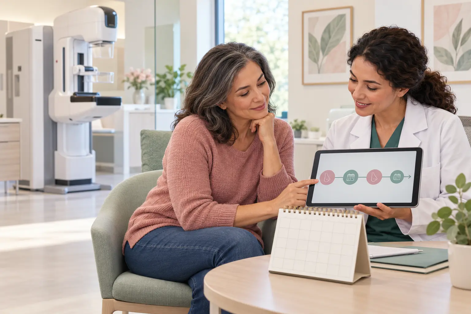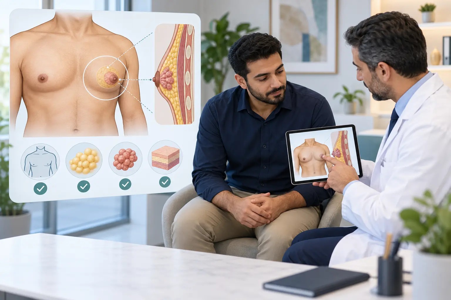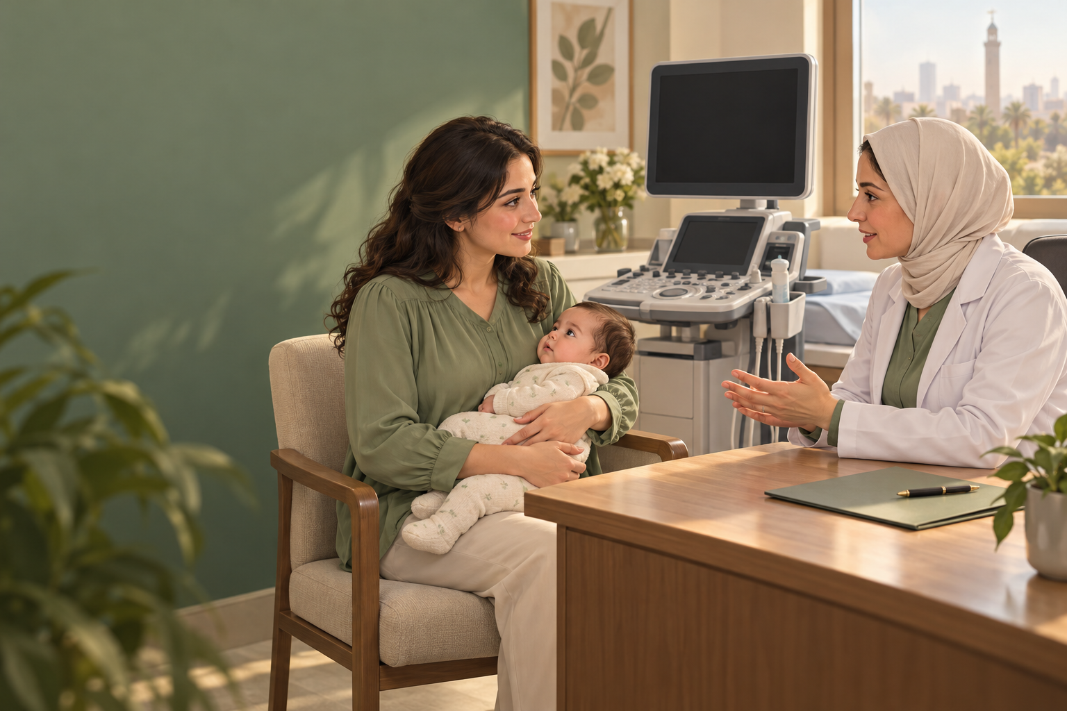
Breastfeeding is not a reason to wait until weaning before investigating a persistent breast lump. Necessary imaging and biopsy can be arranged during lactation. This guide concerns assessment while breastfeeding; pregnancy and cancer-treatment medicines require their own advice. ACR lactation imaging guideline, 2026
Is every lump a blocked milk duct?
No. Milk-retention cysts (galactoceles), lactating adenomas and infection can cause lumps, but unrelated benign or malignant conditions can also occur. Most persistent masses need diagnostic imaging. Arrange assessment for a lump that remains or grows rather than repeatedly assuming milk blockage. Academy of Breastfeeding Medicine, Protocol 30
Which scans can I have while breastfeeding?
Targeted ultrasound and diagnostic mammography, including 3D mammography, have complementary roles. The choice depends on age, symptoms and cancer risk. Lactation does not require postponing these tests. If MRI is indicated, standard gadolinium contrast does not routinely require interrupting breastfeeding. ACR, 2026
Tell the imaging team that you are breastfeeding and whether you could also be pregnant. Feeding or expressing shortly before imaging may improve image quality and comfort. American Society of Breast Surgeons, 2025
When is a core needle biopsy needed?
An image-guided core biopsy obtains tissue under local anaesthetic when a finding needs diagnosis. Breastfeeding can generally continue after the procedure: routine weaning or pumping and discarding milk is not required for the biopsy itself. Ask about any additional medicines or procedures separately. ASBrS oncolactation guidance
What are the biopsy risks? A 2026 evidence update
Discuss bleeding, infection and milk leaking through the skin (a milk fistula). In a retrospective study of 88 lactating patients, 11 developed a fistula and 10 an infection; these groups overlapped. Two needed surgery or hospitalisation, while 9 of the 11 fistulas resolved with continued breastfeeding. This single-centre study does not predict your personal risk. Necessary biopsy should be accompanied by informed consent and aftercare, not a promise of no complications. Langman and colleagues, 2026
What follow-up should I agree before leaving?
Ask when results will be available, whether the sample explains the actual lump and scan, and when reassessment is due. A persistent or growing change needs review even after an initially reassuring assessment. ABM Protocol 30 Read our separate guide to a benign biopsy that does not match suspicious imaging.
While waiting, continue usual feeding if comfortable unless your treating team advises otherwise. Avoid hard massage, extra pumping beyond your baby's needs and abrupt weaning. NHS mastitis advice
Choosing a breast team in Cairo
When searching for “best breast surgeon in Cairo”, use objective criteria: verifiable registration, breast-specific diagnostic experience, access to breast radiology and pathology, and a clear results-and-follow-up plan. The phrase is not an objective ranking. Our surgeon-selection guide explains what to check.
For broader symptom advice, see when a breast lump should be checked. Suspected abscess treatment is covered separately in ultrasound-guided drainage.
Educational information
This is general patient education, not personal medical advice. Your examination, imaging findings and feeding circumstances determine your care. Use the urgent warning signs below rather than waiting for a routine appointment if you become unwell.
Sources checked on 25 October 2026. The six direct references below include professional guidance and a 2026 primary study; older guidance is interpreted alongside the newer evidence.