A fibroadenoma is a benign breast lesion made from glandular and supporting tissue. It is common in younger women, but a new lump at any age still deserves proper assessment. The practical decision is not simply “operation or no operation.” It is whether the finding has been adequately identified, whether the examination, imaging and pathology agree, and whether symptoms, growth or uncertainty make removal worthwhile.
If you have found a new or changing lump, start with the general breast-lump assessment guide. Do not assume that a lump is a fibroadenoma from touch alone.
First ask: how was the fibroadenoma diagnosed?
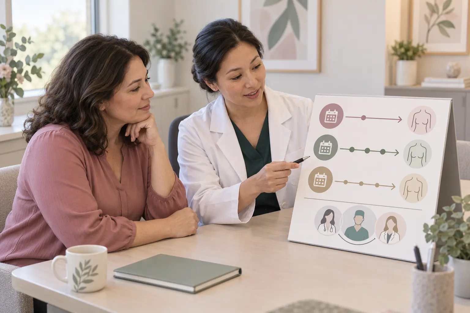
Assessment begins with the history and a breast and lymph-node examination. Imaging is selected for the individual. Under American College of Radiology guidance, ultrasound is usually the first study for a palpable mass in women under 30; ultrasound, diagnostic mammography or tomosynthesis may be appropriate from 30 to 39; and diagnostic mammography or tomosynthesis is generally the starting point from 40, often with targeted ultrasound. Pregnancy, breastfeeding, previous breast disease and personal risk can change the pathway.
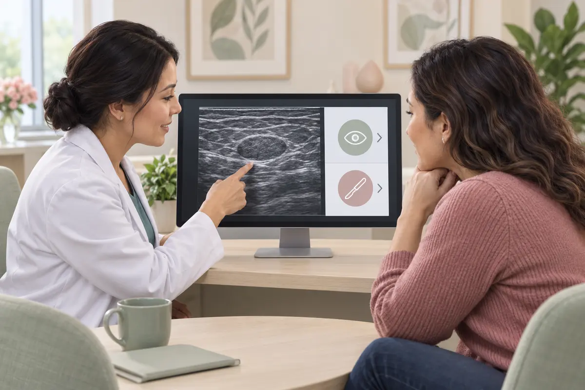
An ultrasound description that looks like a fibroadenoma is not the same as a tissue diagnosis. A radiologist may recommend imaging surveillance for a probably benign finding, or an image-guided core-needle biopsy when the appearance, examination, age, growth or clinical context requires tissue sampling. A core biopsy removes small tissue samples for pathology; it is not the same operation as removing the whole lump.
The key word is concordance. A “concordant” result means the clinical finding, imaging and pathology all tell a compatible benign story. If one part does not fit—for example, a suspicious scan with a benign sample—the result is discordant and needs further review rather than reassurance from the biopsy label alone.
When is observation reasonable?
The 2025 joint guideline from the American Society of Breast Surgeons and Society of Breast Imaging supports observation for a core-biopsy-proven, concordant fibroadenoma without atypia when it is not causing important symptoms, has not shown substantive growth and the patient is comfortable keeping it.
For this specific group, the guideline says lesion-specific imaging follow-up is not required and patients may return to age-appropriate screening. That statement does not apply to a scan-only “probably benign” mass, atypia, radiology–pathology disagreement or concern for a phyllodes tumour. Follow the written recommendation on your own imaging or pathology report; some patients appropriately need short-interval imaging or repeat assessment.
Observation does not mean ignoring the breast. Return earlier if the lump becomes noticeably larger, firmer or more fixed, if new skin or nipple changes appear, or if the symptoms no longer match the original plan.
When should removal be discussed?
Excision may be considered when one or more of the following applies:
- persistent discomfort or pressure attributable to the lump, while recognising that removal does not guarantee that pain will disappear; - substantial or rapid growth, a large lesion that distorts the breast, or increasing uncertainty during follow-up; - atypia, a cellular fibroepithelial lesion, discordant imaging and pathology, or another feature that raises concern for a phyllodes tumour; - a clear patient preference after discussing observation, scars, possible contour or sensation changes, and the chance of another fibroadenoma developing elsewhere.
Size is not a stand-alone diagnosis. The surgeon considers absolute size, change over time, the size of the breast, symptoms, imaging features and pathology together. A stable small lesion with complete concordance is different from a rapidly enlarging mass or an indeterminate fibroepithelial lesion.
Fibroadenoma and phyllodes tumour are not the same
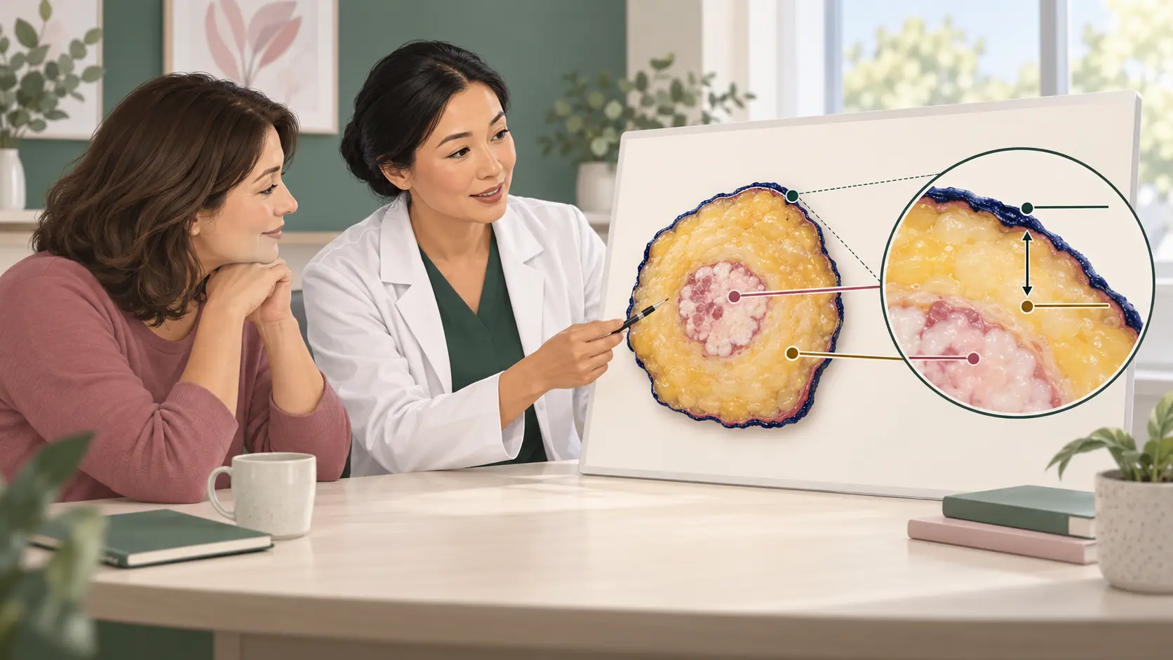
Both fibroadenomas and phyllodes tumours are fibroepithelial lesions, and their appearances can overlap. Many phyllodes tumours are benign, but their behaviour and treatment differ from fibroadenoma. A core sample may occasionally be reported as “fibroepithelial lesion” or “cellular fibroepithelial lesion” because the pathologist cannot confidently separate the two on the sampled tissue.
Rapid or substantive growth does not automatically mean cancer. However, it should trigger timely clinical and imaging reassessment. Depending on the findings, the team may recommend repeat core biopsy or complete excisional biopsy. The latest joint guideline recommends complete excision when pathology or imaging raises concern for a phyllodes tumour.
How can a fibroadenoma be removed?
Open surgical excision removes the lump through a planned incision and sends it for full pathology. When choosing the incision, the surgeon should consider complete removal, breast contour, scar position and preservation of sensation. Review the wider principles in consultant-led planning before breast surgery.
Ultrasound-guided vacuum-assisted excision can remove selected, usually smaller lesions through a small skin opening. The lesion should first be visible on ultrasound, confirmed as a fibroadenoma on core biopsy and concordant with the examination and imaging. Availability and selection criteria vary. Bruising, bleeding, haematoma, incomplete removal and a residual or recurrent lump can occur, so it should not be marketed as risk-free or “scarless.” Cryoablation is another option in some health systems for carefully selected, biopsy-confirmed lesions, but it is not universally available and is not suitable for every location or diagnosis.
After any removal, the tissue result matters. A changed final diagnosis can alter follow-up, so ask how and when you will receive the pathology report.
What is usually expected after biopsy or excision?
Temporary tenderness, bruising, swelling or firmness around the biopsy or operation site can occur. After open excision there will be a scar, and temporary or persistent changes in skin or nipple sensation, a contour dip, bleeding, infection or a fluid collection are possible. Follow the procedure-specific instructions rather than copying another patient’s dressing, activity or medication plan. The breast-surgery wound-healing guide explains normal changes and infection warning signs in more detail.
Red flags and when to seek in-person care
Arrange prompt breast-clinic assessment for a new lump that persists, a previously assessed lump that grows substantially, a hard or fixed mass, skin dimpling, new one-sided nipple inversion, spontaneous bloodstained discharge, or a new underarm lump. These signs do not prove cancer, but they should not be self-diagnosed as fibroadenoma.
Seek same-day urgent assessment for a breast that becomes rapidly swollen, hot, red and painful, especially with fever, chills or feeling unwell; infection or an abscess needs a different pathway.
After biopsy or excision, contact the treating team urgently for a rapidly enlarging or very tense swelling, bleeding that soaks or escapes the dressing, severe or escalating pain, spreading redness, pus or foul-smelling fluid, fever, wound opening, or pale, blue, grey or black skin. Call emergency services for uncontrolled bleeding, fainting or collapse, sudden breathlessness or chest pain, or swelling of the face or tongue with difficulty breathing.
Questions to ask at your appointment
- Is this a probably benign imaging diagnosis or a biopsy-confirmed fibroadenoma? - Do the examination, imaging and pathology agree? - What specific finding makes observation, repeat biopsy, vacuum-assisted excision or open excision appropriate for me? - If we observe it, what change should bring me back and is any scheduled imaging needed? - If it is removed, where will the scar be, what might happen to contour or sensation, and when will pathology be available?
If you search for the best breast surgeon in Cairo, compare objective factors: breast-specific training, documented radiology–pathology concordance, access to image-guided biopsy, a balanced explanation of observation and removal, thoughtful scar planning, full pathology and reliable aftercare. A promotional ranking cannot decide which option is safest for your particular lump.
References reviewed
- Rosenberger et al. — ASBrS/SBI 2025 guideline for benign breast fibroepithelial lesions: https://doi.org/10.1001/jamasurg.2025.4392 - American College of Radiology — Palpable Breast Masses, 2022 update: https://doi.org/10.1016/j.jacr.2023.02.013 - Association of Breast Surgery (UK) — Management of fibroadenomas summary statement: https://associationofbreastsurgery.org.uk/media/h4xne3ie/abs-summary-statement-fibroadenomas-v1.pdf - Dialani et al. — Enlarging biopsy-proven fibroadenoma: is surgical excision necessary?: https://doi.org/10.1016/j.clinimag.2019.03.014 - American Society of Breast Surgeons — 2026 resource guide on ablation and percutaneous excision: https://www.breastsurgeons.org/docs/statements/asbrs-treatment-of-benign-and-malignant-tumors-of-the-breast-2026-02-24.pdf - Giacalone et al. — Outcomes of ultrasound-guided vacuum-assisted excision of fibroadenomas: https://doi.org/10.1007/s11547-023-01684-9
This article is for general patient education only. It does not diagnose a breast lump, interpret your scan or pathology, or replace an in-person examination and an individual plan from your treating team. If a lump is new or changing, or symptoms are severe or worsening quickly, seek licensed in-person medical care rather than waiting for an online reply.